| Hemoglobin | |
|---|---|
 |
|
| General | |
| Abbreviations | Hb or Hgb |
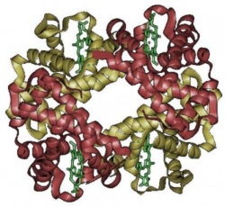
Hemoglobin is a protein that is located in red blood cells, which binds oxygen from the lungs to deliver to the organs and tissues.[1] Hemoglobin is made of two similar proteins, alpha and beta, that are combined together.[2] Since each alpha and beta has its own two protein molecules, Hemoglobin actually consists of four different protein molecules, also called globulin chains. Usually, hemoglobin molecule in the normal adult includes two alpha-globulin chains and two beta-globulin chains. On the other hand, in infants, the hemoglobin molecule contains two alpha chains and two gamma chains. As the infant matures, two gamma chains are changed to two beta chains. Each different globulin chain has the heme molecule, which is iron that carries the oxygen and carbon dioxide in blood. Because of the presence of the heme molecule, blood is a red color. Also, hemoglobin helps to build the shape of the red blood cells. [3] [4]
In 1840, at Leipzig University in Germany, the hemoglobin protein that carries oxygen was discovered by Hünefeld first. In 1851, in his articles about diluting red blood cells with a solvent such as pure water (H2O) or alcohol, Otto Funke, a German physiologist, explained "growing hemoglobin crystals." A few years later, Felix Hoppe-Seyler, a German physiologist and chemist, discovered the binding of oxygen to erythrocytes that is one of hemoglobin's functions. This discovery is also called the process of creating the compound oxyhemoglobin. After studying hemoglobin in crystalline form, Hoppe-Seyler discovered that hemoglobin contains iron. [5] In 1959, using X-ray crystallography, Max Perutz, an Austrian-British molecular biologist, established the molecular structure of hemoglobin. His great discovery helped him win the Nobel Prize for Chemistry in 1962. [6] [7]
Due to problems and mutations in globin gene regulation, there are two kinds of hemoglobins that are produce, normal and abnormal hemoglobins.
One can express the hemoglobin level as the amount of hemoglobin in grams (gm) per deciliter (dl) of blood. (1 deciliter = 100 milliliters)
The normal ranges for Hemoglobin is changeable because of differences in age and the gender of the person.
Usually, a low hemoglobin causes to have anemia. However, there are more causes of low hemoglobin besides anemia;
People who smoke or live at high altitudes can have higher hemoglobin levels than those who do not. Dehydration is the main cause that produces a high hemoglobin. However, unlike other serious diseases, people can lower their high hemoglobin levels easily.
There are more causes that make high hemoglobin levels;
Heme is the prosthetic group of hemoglobin, myoglobin, and the cytochromes. It consists of a porphyrin, known as a heterocyclic ring, is located at the center of the molecule and has an iron atom bound at its center.[9] Globin is the protein that protects the heme molecule from any other dangers. It is also called a globular protein. A single unit of hemoglobin includes a heme that is attached to a globular protein.[10] Hemoglobin synthesis requires the collaboration of heme and globin. In the mitochondria and the cytosol of immature red blood cells, the heme part is combined with a series of steps. On the other hand, the globin protein parts are incorporated with ribosomes in the cytosol. From the proerythroblast to the reticulocyte in the bone marrow, they keep producing hemoglobins in the cell. The nucleus is then removed from mammalian red blood cells. This happens only in humans but not in other species. Although the nucleus is disappeared in red blood cells, residual ribosomal RNA keeps combining and producing hemoglobin until the reticulocyte loses its RNA as soon as it enters the vasculature. In adult humans, hemoglobin is a tetramer, which has two alpha and two beta subunits. The subunits are identical in structure and have equal size. Each subunit has a molecular weight of 16,000 grams. Therefore, a total molecular weight is about 64,000 grams. Since each subunit of hemoglobin includes a single heme, as we aleardy discuss that a heme consists of four different molecules, adult human hemoglobin has a toatla binding capacity of four oxygen molecules. [10]
Some chemical reaction equations:
Therefore,
Besides the binding of hemoglobin for oxygen, the binding of oxygen is also an essential process in the tetrameric form of normal adult hemoglobin. Unlike the normal hyperbolic curve, the oxygen binding curve of hemoglobin is sigmoidal, or 'S' shaped.
However, when carbon dioxide is present and it is at a lower pH level, Hemoglobin's connection to oxygen is decreasing. To get bicarbonate, Carbon dioxide needs to react with water through the reaction:
When the carbon dioxide levels in the blood increase, hemoglobin can combine protons and carbon dioxide, which causes change in the protein and helps to release oxygen. Protons bind a various places with the protein. Also, to form carbamate, carbon dioxide binds at the alpha-amino group. On the other hand, when the carbon dioxide levels in the blood decrease, carbon dioxide is released, increasing the oxygen's connection level of the protein. This, hemoglobin's control of affection of oxygen, is known as the Bohr effect.
Molecules, like 2,3-diphosphoglycerate, which lower the affection of hemoglobin for oxygen, affect the binding of oxygen. When one is used to live in high altitudes, the concentration of 2,3-diphosphoglycerate in the blood is increasing. This allows one to carry a large amount of oxygen to each part of the organs and tissues that have lower oxygen tension. This process is called a heterotropic allosteric effect. [11]
Different hemoglobin variants are due to the abnormal process of producing hemoglobin. Since hemoglobin is made of heme, an iron-containing portion, and globin, which forms a protein, the molecules should be found in all red blood cells. The molecules combine with oxygen in the lungs and carry it around throughout the body to release it to the body’s organs and tissues. [2]
In the embryo:
In the fetus:
In adults:
|
||||||||||||||||||||