|
Tibial plateau fracture Microchapters |
|
Diagnosis |
|---|
|
Treatment |
|
Case Studies |
|
Tibial plateau fracture x ray On the Web |
|
American Roentgen Ray Society Images of Tibial plateau fracture x ray |
|
Risk calculators and risk factors for Tibial plateau fracture x ray |
Editor-In-Chief: C. Michael Gibson, M.S., M.D. [1]; Associate Editor(s)-in-Chief: Rohan A. Bhimani, M.B.B.S., D.N.B., M.Ch.[2]
Overview[edit | edit source]
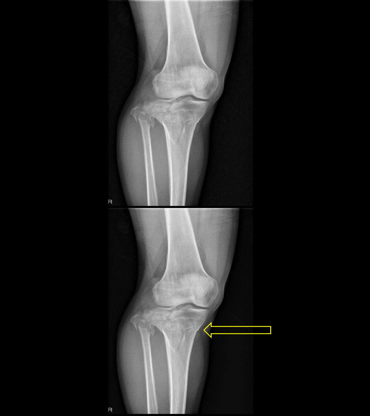
Radiographic imaging is important in diagnosis, classification, treatment and follow-up assessment of tibial plateau fracture. The routine minimal evaluation for tibial plateau fracture must include a antero-posterior (AP) view, oblique and lateral view. The radiological findings include, abnormal joint alignment, depressed articular fragments and coronal split fractures.
X Ray[edit | edit source]
 X ray of Knee showing Schatzker type VI tibial plateau fracture. Source: Case courtesy by: Dr. Rohan A. Bhimani |
- Radiographic imaging is important in diagnosis, classification, treatment and follow-up assessment of tibial plateau fracture.[1]
- The routine minimal evaluation for tibial plateau fracture must include views such as Antero- posterior (AP) view, oblique and lateral view.[1]
- In addition,tibial plateau view which is an AP projection of the knee, angled 15° caudally provides assessment of the depth of plateau surface depression.[2]
- X ray findings include:[3][4][5]
- Antero-posterior view
- Depressed articular surface
- Sclerotic band of bone indicating compression fracture
- Disruption of the joint alignment
- Lateral View
- Posteromedial fracture lines are visualized
- Antero-posterior view
References[edit | edit source]
- ↑ 1.0 1.1 Rossi R, Bonasia DE, Blonna D, Assom M, Castoldi F (2008). "Prospective follow-up of a simple arthroscopic-assisted technique for lateral tibial plateau fractures: results at 5 years". Knee. 15 (5): 378–83. doi:10.1016/j.knee.2008.04.001. PMID 18571417.
- ↑ Schatzker J, McBroom R, Bruce D (1979). "The tibial plateau fracture. The Toronto experience 1968--1975". Clin Orthop Relat Res (138): 94–104. PMID 445923.
- ↑ Markhardt BK, Gross JM, Monu JU (2009). "Schatzker classification of tibial plateau fractures: use of CT and MR imaging improves assessment". Radiographics. 29 (2): 585–97. doi:10.1148/rg.292085078. PMID 19325067.
- ↑ te Stroet MA, Holla M, Biert J, van Kampen A (2011). "The value of a CT scan compared to plain radiographs for the classification and treatment plan in tibial plateau fractures". Emerg Radiol. 18 (4): 279–83. doi:10.1007/s10140-010-0932-5. PMC 3139878. PMID 21394519.
- ↑ Rockwood, Charles (2010). Rockwood and Green's fractures in adults. Philadelphia, PA: Wolters Kluwer Health/Lippincott Williams & Wilkins. ISBN 9781605476773.