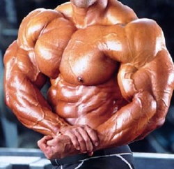
Skeletal muscles are voluntary muscles that help provide movement to the body using contraction and levers. Humans have nearly 620 skeletal muscles that work in concert allowing us coordinated and fluid mobility that is evidence of intelligent design and a testimony to the omniscient creator.[1]
Skeletal muscles are controlled by the central nervous system, and are striated (striped) due to the presence of regularly repeating protein filaments that produce contraction. A whole skeletal muscle (i.e. bicep) is considered an organ of the muscular system. Each organ or muscle consists of skeletal muscle tissue, connective tissue, nerve tissue, and blood or vascular tissue. Skeletal muscles vary considerably in size, shape, and arrangement of fibers. They range from extremely tiny strands such as the stapedium muscle of the middle ear to large masses such as the muscles of the thigh. Some skeletal muscles are broad in shape and some narrow. In some muscles the fibers are parallel to the long axis of the muscle; in some they converge to a narrow attachment; and in some they are oblique.[2]
Each skeletal muscle cell is long and cylindrical and often called a muscle fiber. An individual skeletal muscle may be made up of hundreds, or even thousands, of muscle fibers bundled together and wrapped in a connective tissue covering. Each muscle is surrounded by a connective tissue sheath called the epimysium. Fascia, connective tissue outside the epimysium, surrounds and separates the muscles. Portions of the epimysium project inward to divide the muscle into compartments. Each compartment contains a bundle of muscle fibers. Each bundle of muscle fiber is called a fasciculus and is surrounded by a layer of connective tissue called the perimysium. Within the fasciculus, each individual muscle cell, called a muscle fiber, is surrounded by connective tissue called the endomysium.[2]
Skeletal muscle cells (fibers), like other body cells, are soft and fragile. The connective tissue covering furnish support and protection for the delicate cells and allow them to withstand the forces of contraction. The coverings also provide pathways for the passage of blood vessels and nerves.[2]
Commonly, the epimysium, perimysium, and endomysium extend beyond the fleshy part of the muscle, the belly or gaster, to form a thick ropelike tendon or a broad, flat sheet-like aponeurosis. The tendon and aponeurosis form indirect attachments from muscles to the periosteum of bones or to the connective tissue of other muscles. Typically a muscle spans a joint and is attached to bones by tendons at both ends. One of the bones remains relatively fixed or stable while the other end moves as a result of muscle contraction.[2]
Skeletal muscles have an abundant supply of blood vessels and nerves. This is directly related to the primary function of skeletal muscle, contraction. Before a skeletal muscle fiber can contract, it has to receive an impulse from a nerve cell. Generally, an artery and at least one vein accompany each nerve that penetrates the epimysium of a skeletal muscle. Branches of the nerve and blood vessels follow the connective tissue components of the muscle of a nerve cell and with one or more minute blood vessels called capillaries.[2]
Each skeletal muscle is attached to two bones: one stationary bone and one bone that moves greatly. There are three parts to a muscle that is attached to these bones. They are called, in relation to skeletal muscle, the origin,the insertion, and the belly. The origin is where a muscle's tendon is connected to a relatively stationary bone. The insertion is where a muscle's tendon is connected to a relatively mobile bone. The belly is the part of the muscle that contains muscle cells. It is also the largest part of a skeletal muscle, and is located between the origin and insertion.[3]
Skeletal muscle is controlled by the Central nervous system (CNS) and is used for skeletal movements. Skeletal muscles are the only voluntary muscles in the body and they are striated. Skeletal muscles use the bones they are connected to as levers and contraction as a means to move.[4]
Bones are used for movement, but are not able to move by themselves. They require the use of contraction and relaxation of skeletal muscles.[5] In order to move in the human body, skeletal muscles must make use of levers. Skeletal muscles create pull on bones and use them as levers. Levers are moved by a force referred to as an effort. The area to which this force is applied is referred to as a point of effort. The force to be moved is referred to as a load or resistance. The distance between the fulcrum and the point of effort is referred to as a load arm.[5] There are three types of levers found in the muscle system: the first-class lever, the second-class lever, and the third-class lever. In the first-class lever the fulcrum resides in between the resistance and effort.[6] An example of a first-class is seen when a bent arm is made straight. Another example a first-class lever is seen in the body in the head when it goes in a nodding motion.[5] The next type of lever seen in the human body is the second-class lever. In the second-class lever, the resistance is in the middle of the fulcrum and effort.[7] This lever is seen in the foot when walking. In the third-class lever the effort resides in between the resistance and fulcrum. An example of this is found in the bicep when it contracts while lifting an object.[5]
Bones have no means of movement on their own, so to move, they use the contraction and relaxation of skeletal muscles.[5]There are four different kinds of contractions. These contractions are: Concentric contractions, eccentric contractions, isometric contraction, and passive stretch. Concentric contractions are contractions that permit muscles to shorten less than their max tetanic tension. A concentric contraction can be seen when you do a bicep curl with a weight. Eccentric contractions are seen when load on a muscle reaches a point at which outside force is more than the force a muscle can generate and are forced to become extended because of a high external load. Isometric contraction is seen when a muscle is activated, but rather than extending or shortening it is kept at an unchanging length. An example of this type of contraction is seen when you carry a load in front of you. The last type of contraction is passive stretch. This is when a muscle is extended during it's passive state. You can see an example of this when you touch your toes while feeling your hamstrings.[8]
Myopathy: Myopathies are neuromuscular diseases where dysfunction of muscle fibers cause muscle weakness. Muscle weakness is the primary symptom but other symptoms may include muscle spasms, muscle stiffness, and muscle cramps. Myopathies can be genetical or may be transmitted. There are several groups of myopathies. They can be grouped as congenital myopathies, myoglobinurias, glycogen storage diseases of muscle, muscular dystrophies, mitochondrial myopathies, dermatomyositis, myositis ossificans, familial periodic paralysis, polymyositis, inclusion body myositis, and related myopathies, neuromyotonia, stiff-man syndrome, common muscle cramps and stiffness, or tetany.[9]
Chronic Fatigue Syndrome: Chronic fatigue syndrome is a disease with symptoms of fatigue, headache, muscle pains, chronic sore throats, and concentration difficulties.[10] Other symptoms include chest pain, recurring cough, irritable bowels, nausea, and jaw pains. [11]
Polymyositis: Polymyositis is a connective tissue disease associated with inflamed muscles. This disease leads to muscle weakness and muscle degeneration. The effect of this weakness is experienced greatest in muscles that are near your trunk, in the shoulder muscles or hip muscles, for example. Polymyositis can be seen by people of all ages, but is most evident in adults of about age 40 or 50. Polymyositis is seen more often in women than men and is most commonly in blacks.[12]
Dermatomyositis:
Dermatomyositis a myopathy characterized by skin rash and inflammation. There is no known cause to dermatomyositis, but researches believe that it may be an infection caused by a virus. They also think that the cause may be a problem in the body's immune system. This disease has the same characteristics of polymyositis, but polymyositis does not have a skin rash associated with it. Dermatomyositis can be seen in many different kinds of people, but is most often seen in children of ages 5 through 15. Most adults seen with it range from age 40 through 60. Dermatomyositis is also most often seen in women rather than men. Symptoms for dermatomyositis include trouble swallowing, red or purple skin rash, violet hued upper eyelids, weakness, stiffness, or soreness of muscles, and difficulty breathing. [13]
Muscular dystrophy: Muscular dystrophies are a group of genetic diseases which are marked by skeletal muscles that have increasing weakness and atrophy of skeletal muscles. The skeletal muscles affected are those that influence movement. There are over thirty genetic diseases associated with this group. Muscular dystrophy may be seen in infants or children. Other cases aren't seen until adult ages or even later. Duchenne muscular dystrophy most often affects boys and is the most seen muscular dystrophy. Duchenne muscular dystrophy is the result of a lack of dystrophin. Another form of muscular dystrophy is facioscapulohumeral muscular dystrophy. This type of muscular dystrophy most often begins in years of adolescence. Facioscapulohumeral muscular dystrophy results in steady weakness of many muscles of the body. Another type of major muscular dystrophy is myotonic muscular dystrophy. This type of muscular dystrophy is found most commonly in the adult years. This type of muscular dystrophy is characterized extended muscle spasms, cardiac anomalies, cataracts, and endocrine agitations.[14]
Fibromyalgia: Fibromyalgia is a muscle disease that causes pain in the muscles. Fibromyalgia also results in tender areas that result in pain when touched. Fibromyalgia symptoms include difficulty sleeping, headaches, stiffness in the morning, and difficulty with memory and thinking. The cause of fibromyalgia is currently unknown. It is most often seen in middle aged women, but any body can contract it.[15]
Rhabdomyolysis: Rhabdomyolysis is a disease that causes the collapse of muscle fibers. This failure results in the discharge of myoglobin into the blood flow. Symptoms include abnormally colored urine, overall weakness, muscle stiffness or tenderness, and muscle weakness. The cause of the disorder may be caused by damage or trauma to skeletal muscle.[16]